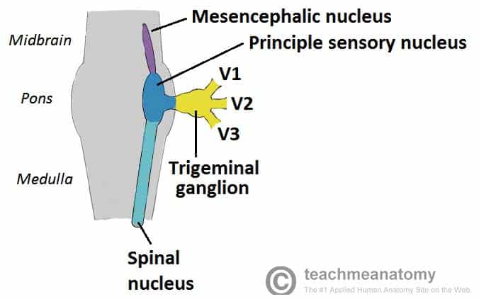
The trigeminal nerve (Cranial Nerve V) is responsible for sensation in the face and certain motor functions like biting and chewing. It has both sensory and motor components, and these are organized in different structures: the sensory ganglion and the motor fibers.
Here’s a breakdown of the key differences:
1. Trigeminal Ganglion (Sensory)
The trigeminal ganglion is a collection of nerve cell bodies (a ganglion) that serves as the primary sensory ganglion for the trigeminal nerve. It is located in the middle cranial fossa, within a bony cavity called the Meckel’s cave.
- Function: It carries sensory information from the face, including touch, pain, temperature, and proprioception.
- Divisions: The trigeminal nerve splits into three major sensory branches after the ganglion:
- Ophthalmic (V1): Sensory for the forehead, eyes, and scalp.
- Maxillary (V2): Sensory for the cheek, upper jaw, and upper teeth.
- Mandibular (V3): Sensory for the lower jaw, lower teeth, chin, and part of the tongue.
- Structure: The ganglion contains the cell bodies of the sensory neurons. The axons of these sensory neurons pass from the ganglion to the sensory nuclei in the brainstem, where the sensory signals are processed.
2. Motor Component of Trigeminal Nerve
The motor component of the trigeminal nerve is responsible for the movement of muscles involved in chewing (mastication). The motor fibers arise from a nucleus in the brainstem called the motor nucleus of the trigeminal nerve, which is located in the pons.
- Function: The motor fibers control the muscles of mastication (e.g., masseter, temporalis, medial and lateral pterygoid muscles) and some additional muscles, including the mylohyoid and tensor tympani (involved in hearing).
- Motor Pathway: The motor fibers of the trigeminal nerve are bundled within the mandibular division (V3) of the trigeminal nerve, even though the motor nucleus is separate from the sensory ganglion. The motor fibers exit the brainstem and travel to the muscles they innervate.
Key Differences:
| Aspect | Trigeminal Ganglion (Sensory) | Motor Component of Trigeminal Nerve |
|---|---|---|
| Function | Sensory information (touch, pain, etc.) | Motor control of chewing muscles |
| Location | Meckel’s cave (Middle cranial fossa) | Pons (Brainstem) |
| Innervates | Sensory regions of the face | Muscles of mastication and others (e.g., tensor tympani) |
| Pathways | Sensory fibers travel to brainstem for processing | Motor fibers travel to muscles via mandibular division (V3) |
| Neurons | Contains cell bodies of sensory neurons | Motor neurons originate in the pons and synapse in muscles |
Summary:
- The trigeminal ganglion is the sensory hub, collecting sensory input from the face and transmitting it to the brainstem.
- The motor component of the trigeminal nerve controls the muscles used in chewing, and these fibers travel along the mandibular division of the nerve. Both sensory and motor functions are crucial for proper facial sensation and mastication, but they involve different anatomical structures and pathways.
